Research Article 
 Creative Commons, CC-BY
Segment Transport with Ilizarov that Causes Spontaneous Healing of Gap at Docking Site in Long Bones, a Case Series and Review of Literature
*Corresponding author: Imran Khan, Assistant Professor, Department of Orthopedic and Trauma Medical Teaching Institute Lady Reading Hospital Peshawar Pakistan.
Received: April 06, 2023; Published: April 12, 2023
DOI: 10.34297/AJBSR.2023.18.002481
Summary
Healing of bone is dependent on multiple factors but the most common one is when the ends are in fully contact, and the fracture is close. This ideal situation does not occur when there is an open fracture due to motor vehicle accident or firearm injuries. The management of such types of fractures is always challenging for the orthopedic surgeons [1]. While managing open fracture the orthopedic surgeon does wound excision and sometime remove the dead bone from fracture ends that leads to formation of gap at fracture site. Theoretically the gap may not heal without intervention [2]. The surgical intervention for filling the gap may either be cancellous bone graft, strut graft, cortical graft, intramembranous technique, with segment transport or shortening of the bone [3,4] For large gap, cortical graft, like fibular graft, or segment transport are widely used procedures which are technical demanding and time-consuming procedures [5]. Lazaro has the advantage of eradicating the infection while at the same time to fill the gap with segment transport. Segment transport may be bifocal, trifocal or tetra focal depending on the condition of the bone and size of the gap [6]. This case series of three patients have unique bone healing at the fracture site while transporting segment and the gape was ultimate filled without any intervention. All the cases have fracture due to trauma and were initially fixed internally and later developed infection. All patients have no head injury at initial impact.
Case No 1
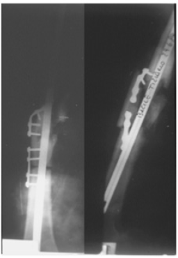
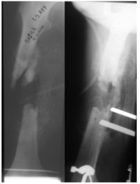
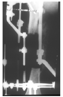
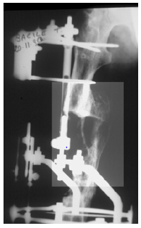
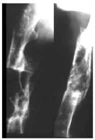
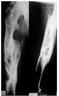
A male, age 18 years had a road traffic accident and sustained multiple injuries. In spite of other injuries was diagnosed as the fracture of femur, clavicle and ribs. He was managed with internal fixation, but it became heavily infected in another hospital. Then he was shifted to our unit for management. As was very weak and had chest problem due to rib fracture so he was initially admitted in intensive care unit. Once he became stable then we managed him with removal of implants and did debridement, but the infection was not controlled. Then in the second stage we removed the dead ends of fracture site that has created created a gap of 11centimeters. To stabilize the bone we applied Lazaro for control of infection and to optimize the patient. At this stage there was no osteotomy done due to the health condition of the patient. The patient was discharged and advised for regular follow up. The patient was very cooperative and very keen in his health. He visited the Lazaro clinic regularly for control of infection. While we were waiting for optimization of his health, we found callus in the gap. We followed him up for six months in Lazaro and found that all the of gap was filled with new bone and healed without any intervention (Figures 1-6).
Case No 2
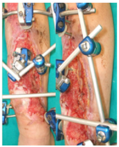
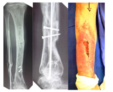
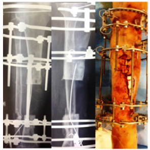
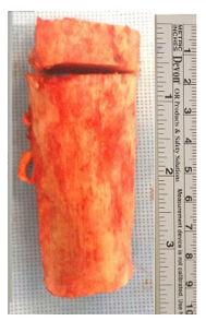
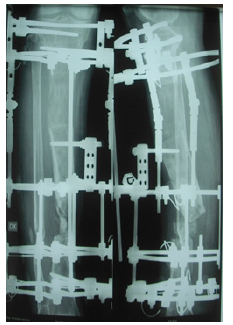
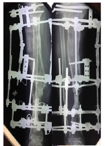
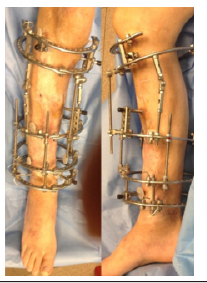
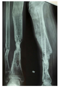
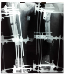
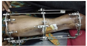
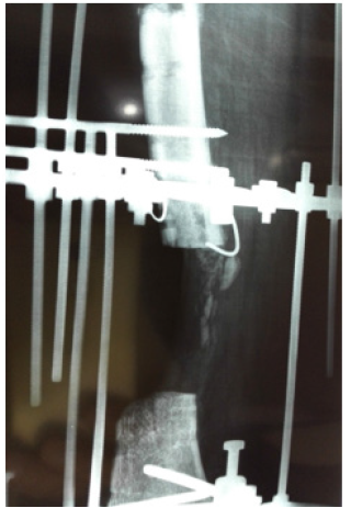
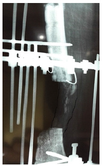
A male aged 60 had motor vehicle accident and sustained open fracture (Gustilo and Anderson type 3 b) of the tibia. Initially, he managed in another hospital with unplaster external fixator with screws in tibia and Kirschner wire in fibula. As there was skin loss so flap was applied over it. A few days late the flap was failed and there was pouring pus coming out of wound. In the first step he was referred to infective medicine unit of our hospital for infection control. A lot of antibiotics has been tried but insane. They have requested our unit for help in managing to eradicate infection. All antibiotics were stopped debridement with wound excision was done but it did not heal. Then all the implants were removed and daily dressing was advised to control infection but no improvement. Finally a resection of the nine centimeter of infected segment of bone done with application of Ilizarov fixator proximal osteotomy for bone transport. No antibiotics were used in this procedure. Skin loss were coverd without any procedure after application of Ilizarov that controlled the infection. With Ilizarov there is darmatotaxis which cover the skin loss. After few follow ups of bone transport the patient felt severe pain when he rotated the nuts. On examining the patient, everything was found normal except the lateral radiograph, which showed the transporting bone was deviated from its path. Deviation was corred with horizontal rods while the transport was stopped. In the next follow up visit we saw new bone formation within the gap. We stoped everything for the gap to be healed spontaneaouly without transport. In this patient, a 5 centimeter bone has been transported (Figures 7-12).
A fifty years old smoker had a history of motor vehicle accident and fracture of femur that was fixed with plate and screws. The plate was broken and was removed and intramedullary nail was put in with iliac crest bone graft. It became infected and was removed. Then uniplanar external fixator was applied but neither infection control nor union achieved. After three years of the initial trauma the patient again present to us for the same problem. Initially we stopped all medicines and stressed the patient for cession of smoking. After three months of cession of smoking, We applied Ilizarov fixator did proximal corticotomy for transport and removed the dead ends till bleeding ends which created a gap of 6 centimeter. After one month of transport there was sever pain while rotating the nuts. When we did radiographs, we saw callus formation in front of transport segment. we stopped the transport and advise the patient for active exercises and regular follow up at one month interval after six month the gap was filled and bone healed without any intervention.
Discussion
Gap and infected non-union is a real challenge for orthopedic surgeons [5,7]. In infected non-union, most of the times, bone endings are cut short a long with debridement of soft tissues in order to eliminate the infection [8]. The gap may be filled with different procedures. If the gap is less than one centimeter then either end to end contact of fragment should be done with shortening of the limb or bone graft may be put in to fill the gap [9]. If the gap is large then it may be filled with vascularized fibular graft-which is a lengthy procedure and a hilarious, technical demanding job [10]- or with bone transport [11]. Removal of fibula for grafting also disturbed the normal mechanic of lower limb which created problems in knee and ankle joints [12]. The gap may also be filled and treated with intra-membranous new bone formation but this is a new technique and needs a lot of cancelleous bone to be put in the gap [13]. Bone transport may be done either with monoliteral external fixator or with circular ring fixator [14]. The circular ring fixator which is commonly known as Lazaro apparatus, nowadays in vogue throughout the world. The application Felizardo fixator is also not an easy procedure, it is also technical demanding for the surgeon as well as for the patient. After application, care of the pins, frequent visits to the clinic and daily adjustment to the nuts for lengthening is not an easy practice [15]. Secondary procedure in Ilizarov fixator are common and docking site needs almost always reoperation.
We also tried the Ilizarov fixator in both of these cases but due to spontaneous healing of the gap bone transport was abandoned. There are other therapeutic agents that are sometimes used to hasten the union but none of them was used in these two cases.
Until now: thirty-two cases of spontaneous regeneration of bone loss in mandible [16], one in humerous, two in tibia, one in fibula and five case of femur has been reported in the literature. The first case of bone regeneration in tibia, after resection in osteomylitis, was noted by Cheever in 1870.17 After that a lot of study was done by Xichols in 1902 and Phemister in 1915.17Klein DM et al7 reported a case with 20 centimeter bone loss in tibia that has been healed spontaneously. Bosworth DM, et al [17] study shows six patients with tibial defects that has been spontaneously regenerated and healed. Park HW, et al [18] reported a three years old boy with road traffic accident in which he lost whole of lateral malleolus and was regenerated fully in four years time. Hinsche AF, et al [19] in 2003, study has noted spontaneous regeneration of gap 6, 9, 10 and 15 centimeters in femur in four patients. Kiter E et al [20] in 2010, noted one patient with huge gap in femur while Pazzaglia UE, et al [21] in 1991, has also noted one patient with loss of proximal femur that healed spontaneously in eight years old child. Ulkü O, et al. [22] has reported a case in 1997 with spontaneous bone regeneration of 16 centimeter in distal humerus which was lost due to agricultural machine injury.
What strike mind that for bone healing to occur, the bone ends must be in close contact or there may be some other forces or factors that heals the bone. These forces or factors may be supernatural, genetic makeup of the patient or local environment at fracture site that heals the gap which we do not know till date. Research is needed to find out that forces or factors, which will make easier the management of gap in bones.
Acknowledgments
None.
Conflict of Interest
None.
References
- Webb JCJ, Tricker J (2000) A review of fracture healing. Curr Orthop 14(6): 457-463.
- Packer JW, Colditz JC (1986) Bone injuries: treatment and rehabilitation. Hand Clin 2(1): 81-91.
- Oryan A, Alidadi S, Moshiri A (2013) Current concerns regarding healing of bone defects. Hard Tissue 2(2): 13.
- Ilizarov GA (1988) The principles of the Ilizarov method. Bull Hosp Joint Dis Orthop Inst 48: 1-11.
- Guerreschi F, Azzam W, Camagni M, Lovisetti L, Catagni MA (2010) Tetrafocal Bone Transport of the Tibia with Circular External Fixation; A Case Report. J Bone Joint Surg Am 92(1): 190 -195.
- Smith WR, Elbatrawy YA, Andreassen GS, Philips GC, Guerreschi F, et al. (2007) Treatment of traumatic forearm bone loss with Ilizarov ring fixation and bone transport. International Orthopaedics (SICOT) 31: 165-170
- Klein DM, Caligiuri DA, Riina J, Katzman BM (1997) Spontaneous healing of a massive tibial cortical defect. J Orthop Trauma 11(2): 133-135.
- Enneking WF, Eady JL, Burchardt H (1980) Autogenous cortical bone grafts in the reconstruction of segmental skeletal defects. J Bone Joint Surg Am 62(7): 1039-1058.
- Chen ZW, Chen LE, Zhang GJ, Yu HL (1986) Treatment of tibial defect with vascularized osteocutaneous pedicled transfer of fibula. J Reconstr Microsurg 2(3): 199-205.
- Newington DP, Sykes PJ (1991) The versatility of the free fibula flap in the management of traumatic long bone defects. Injury 22(4): 275-281.
- Aronson J, Johnson E, Harp JH (1989) Local bone transportation for treatment of intercalary defects by the Ilizarov technique: biomechanical and clinical considerations. Clin Orthop 243: 71-79.
- Ilizarov GA, Ledyaev VI (1992) The Replacement of long tubular bone defects by lengthening distraction osteotomy of one of the fragments. Clin Orthop Related Res 280: 7-10.
- Pelissier P, Masquelet AC, Bareille R, Pelissier SM, Amedee J (2004) Induced membranes secrete growth factors including vascular and osteoinductive factors and could stimulate bone regeneration. J Orthop Res 22(1): 73-79.
- Catagni MA, Guerreschi F, Cattaneo R (1999) Treatment of infected nonunion with the Ilizarov method. Giornale Italiano di Ortopedia e Traumatologia. 24: 443-451.
- Catagni MA, Guerreschi F, Lovisetti L (2011) Distraction osteogenesis for bone repair in the 21st century: lesson learned. Injury 42(6): 580-586.
- Ahmad O, Omami G (2015) Self-regeneration of the mandible following hemi-mandibulectomy for ameloblastoma: a case report and review of literature. J Maxillofac Oral Surg 14(Suppl 1): 245-250.
- Bosworth DM, Liebler WA, Nastasi AA, Hamada K (1966) Resection of the tibial shaft for osteomyelitis in children. A thirty two year follow-up study. J Bone Joint Surg Am 48(7): 1328-1339.
- Park HW, Kim HJ, Park BM (1997) Spontaneous regeneration of the lateral malleolus after traumatic loss in a three-year-old boy: a case report with seven-year follow-up. J Bone Joint Surg Br 79(1): 66-67.
- Hinsche AF, Giannoudis PV, Matthews SE, Smith RM (2003) Spontaneous healing of large femoral cortical bone defects: does genetic predisposition play a role? Acta Orthop Belg 69(5): 441-446.
- Kiter E, Akkaya S, Oto M, Günal I (2010) Spontaneous regeneration of the large femoral defect in patient with diffuse osteomyelitis after intramedullary nailing. Eklem Hastalik Cerrahisi 21(3): 178-181.
- Pazzaglia UE, Finardi E, Pedrotti L, Zatti G (1991) Fracture with loss of the proximal femur in a child. A case report. Int Orthop 15(2): 143-144.
- Ulkü O, Karatosun V (1997) Regeneration of bone after loss of the distal half of the humerus. Case report with a 20-year follow-up. J Bone Joint Surg Br 79(5): 746-747.



 We use cookies to ensure you get the best experience on our website.